X-ray detection
Scintillation materials used in X-ray detectors require high X-ray absorption efficiency and light output, low afterglow, transparency, and spectral response matched to photoelectric converters. The performance of different scintillators varies greatly. In order to be used with a specific CCD, we should consider the luminescence efficiency, spectral response and other characteristics, and select the appropriate scintillation material.
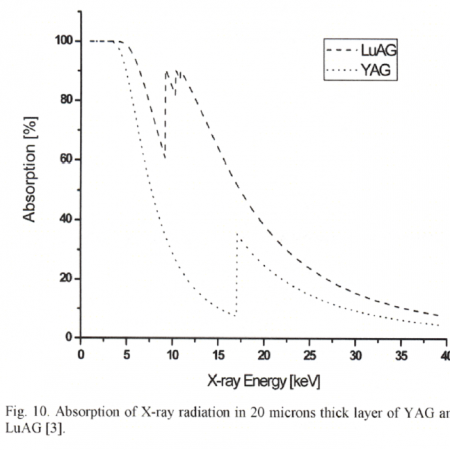
The average absorption depth of X-ray radiation in scintillators depends on the photon energy and material. The advantage of material transparency decreases with the thickness of the imaging plate. If the scintillator is thinner, the average absorption depth will be lower and the resulting image will be sharper due to less image blurring due to less lateral spread of the scintillation photons. Therefore, the thinner the imaging plate, the better the resolution obtained in the image. On the other hand, the detection efficiency decreases with the thickness of scintillator.
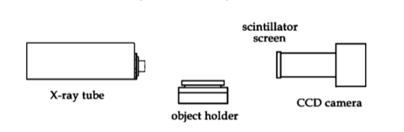
The X-ray detector based on CCD is composed of scintillator, optical coupler and CCD array. It covers a thin layer of scintillators on a linear CCD array. The scintillator converts the X-rays into visible light sensitive to the CCD, which is then converted into an electrical signal reflecting the distribution of the X-rays in space.
Scintillation crystals for X-ray detection
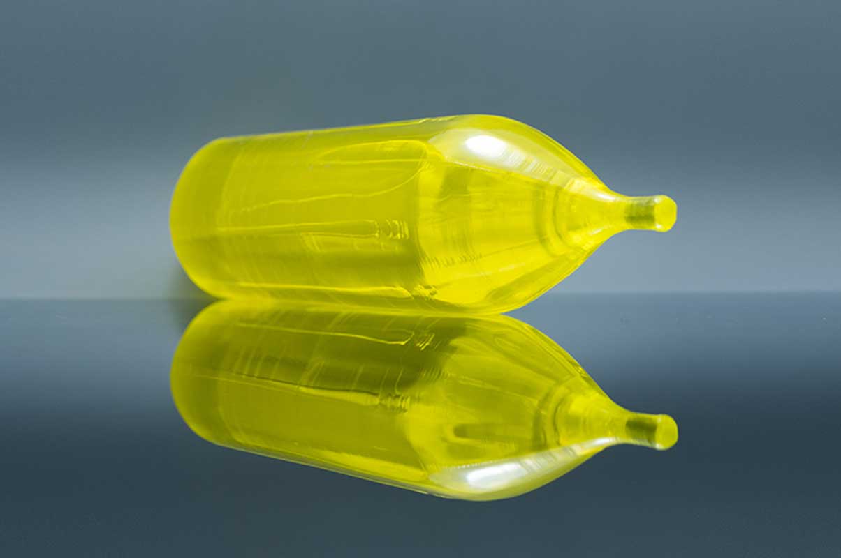
Ce:YAG
| Wavelength (Max. Emission) (nm) | 550 |
| Decay time (ns) | 70 |
| Light yield (Photons/kev) | 35 |
| Refractive index | 1.82@550nm |
| Radiation length (cm) | 3.5 |
Ce:YAG is a fast scintillator with excellent mechanical and chemical tolerance. Its decay time is about 70 ns, which enables high count rates, and the quantum yield is estimated to be between 40 and 50 pH/keV, although the light output is small. However, its non-hygroscopicity and short decay time are still superior to the standard CsI:Tl scintillation, and the maximum emission wavelength (about 550 nm) is very suitable for the sensitivity of the photocathode of the PMT module. Experiments show that Ce:YAG and Ce:LuAG screens are suitable for high spatial resolution imaging. The resolution of the proposed imaging system is about 1 micron.
References
[1] An X-ray counting system based on YAG Ce scintillator
[2] Thin YAG:Ce and LuAG:Ce single crystal imaging plates used for high spatial resolution in X-ray imaging systems
[3] Similarity of trap state and thermoluminescence processes of Ce YAG for X-ray and UV irradiation
[4] High-resolution imaging of biological and other objects with an X-ray digital camera
[5] High-resolution application of YAG Ce and LuAG Ce imaging detectors with a CCD X-ray camera
Ce:YAP
| Wavelength (Max. Emission) (nm) | 370 |
| Decay time (ns) | 28 |
| Light yield (Photons/kev) | 25 |
| Light output relative to Nal (TI) (%) | 60-70 |
| Refractive index | 1.95@370nm |
The characteristics of Ce:YAP crystals as excellent scintillators include: (I) a fast decay time of 30 ns, (ii) a high density of 7.4 g/cm3, and (iii) a high light yield of approximately 30% in NaI (Tl) scintillators. Ce is a fast emitting scintillator, mainly used in PET and animal PET detectors.
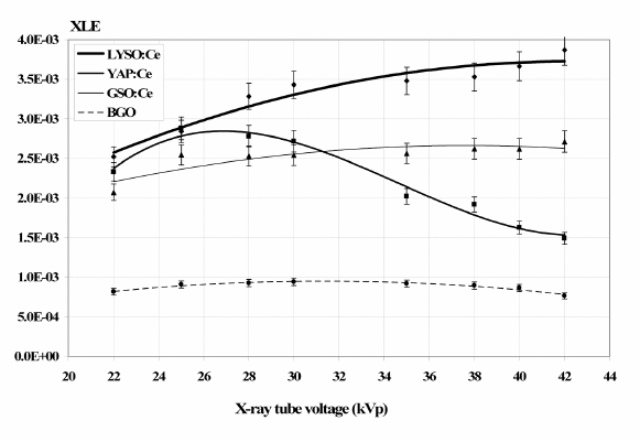
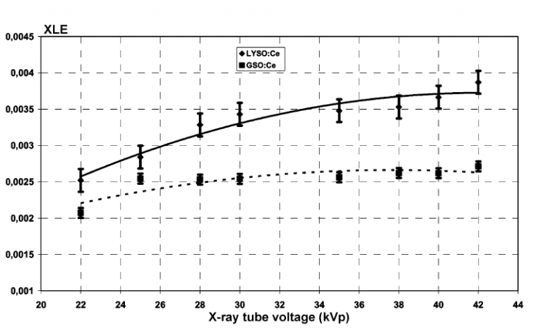
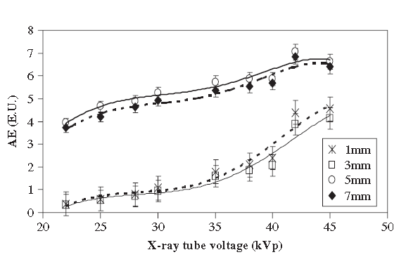
The x-ray luminescence efficiency (XLE) of Ce:LYSO, Ce:YAP, Ce:GSO and BGO as determined by the experimental data for x-ray tube voltages between 22–42kVp(mammography).Points: measured data, line: fitted curve.
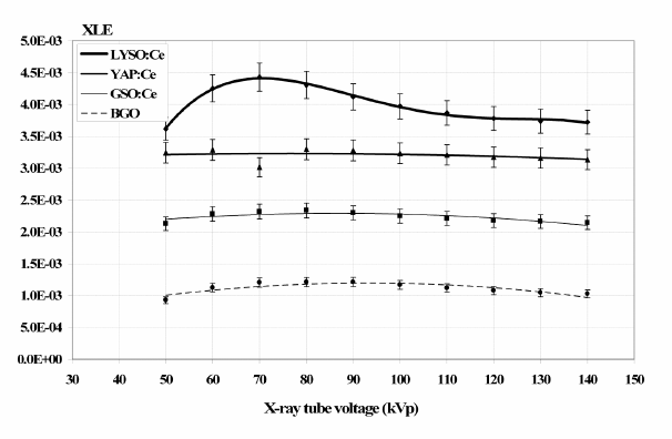
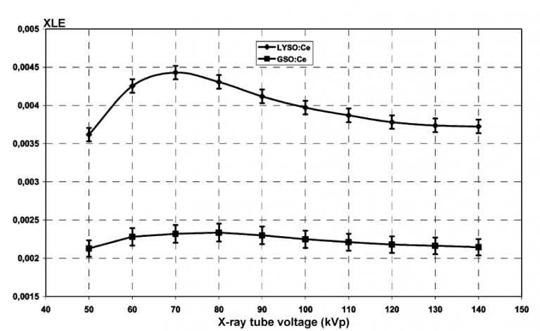
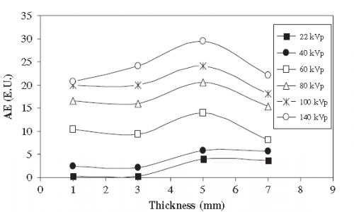
The x-ray luminescence efficieny (XLE) of Ce:LYSO, Ce:YAP, Ce:GSO and BGO as determined by the experimental data for x-ray tube voltages between 50–140 kVp (general radiography).Points: measured data, line: fitted curve.
References
[1] A YAP(Ce) imager operated in high energy X-ray region
[2] Comparative evaluation of single crystal scintillators under x-ray imaging conditions
[3] Comparative Investigation of Ce Doped Scintillators in a Wide Range of Photon Energies Covering X-ray CT, Nuclear Medicine and Megavoltage Radiation Therapy Portal Imaging Applications
[4] ISPA tubes with scintillating YAP Ce windows X- and γ- ray imaging
[5] High-resolution application of YAG Ce and LuAG Ce imaging detectors with a CCD X-ray cameraMcps-range photon-counting X-ray computed tomography system utilizing an oscillating linear-YAP(Ce) photon detector
[6] Properties of a YAP Ce detector for high-energy X-ray counting experiments
Ce:LYSO

| Wavelength (Max. Emission) (nm) | 410 |
| Decay time (ns) | 40 |
| Light yield (Photons/kev) | 25 |
| Light output relative to Nal (TI) (%) | 75 |
| Radiation length (cm) | 1.82@410nm |
Lutetium – yttrium-based scintillators, such as Ce:LYSO, have high effective atomic numbers, are non-hygroscopic, fast emitting materials, and are promising for positron emission imagers.
Ce:LYSO is a hybrid LSO/YSO (5-10%) non-hygroscopic crystal with high density (7.1 g/cm3), high light output (30000 pH/MeV), good energy resolution (10%) and short decay time (40 ns). Although Ce:LYSO and Ce:LSO exhibit similar behavior, with respect to the attenuation scheme, Ce:LYSO appears to have approximately 20% higher light yield than LSO under low-energy (35 kV) X-ray excitation.
References
[1] A comparative study of the luminescence properties of LYSO Ce, LSO Ce, GSO Ce and BGO single crystal scintillators for use in medical X-ray imaging
[2] Comparative evaluation of single crystal scintillators under x-ray imaging conditions
[3] Comparative Investigation of Ce Doped Scintillators in a Wide Range of Photon Energies Covering X-ray CT, Nuclear Medicine and Megavoltage Radiation Therapy Portal Imaging Applications
[4] Evaluation of the light emission efficiency of LYSO Ce scintillator under X-ray excitation for possible applications in medical imaging
[5] Improving Ce3+ doped scintillating materials for medical imaging applications
[6] Investigation of luminescence emission properties of LYSO Ce and LuYAP Ce single crystal scintillators under x-ray exposure for use in medical imaging
[7] Luminescence Properties of LYSO ce and GSO Ce Single Crystal Scintillators Under X-Ray Excitation for Use in Medical Imaging Systems
[8] Scintillator efficiency study with MeV x-rays
Ce:GAGG
| Wavelength (Max. Emission) (nm) | 520 |
| Wavelength range (nm) | 475-800 |
| Decay time (ns) | 90 |
| Light yield (Photons/kev) | 50 |
| Refractive index | 1.95@540nm |
Ce:GAGG (Ce:Gd3Al2Ga3O12) scintillation crystal with high density (6.69 g/cm3), fast scintillation response (~ 90 ns) and high optical yield (~ 46,000 pH/MeV). Ce:GAGG is non-hygroscopic (e.g., Ce:LaBr3, Ce:LaCl3, CsI, and NaI:Tl crystals) and does not have natural radioactivity (e.g., based materials) in contrast to many current highly efficient scintillators. Ce:GAGG emits photons at 520nm with an effective atomic number equal to 54.4.
References
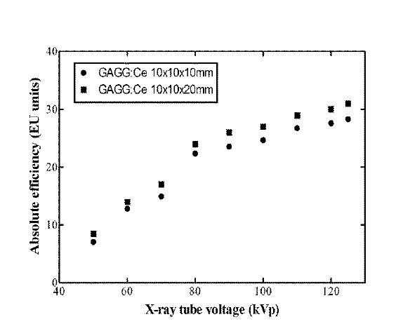
[1] X-ray Luminescence Efficiency of GAGG Ce Single Crystal Scintillators for use in Tomographic Medical Imaging System
[2] Light Emission Efficiency of GAGG Ce Single Crystal Under X-ray Radiographic Conditions
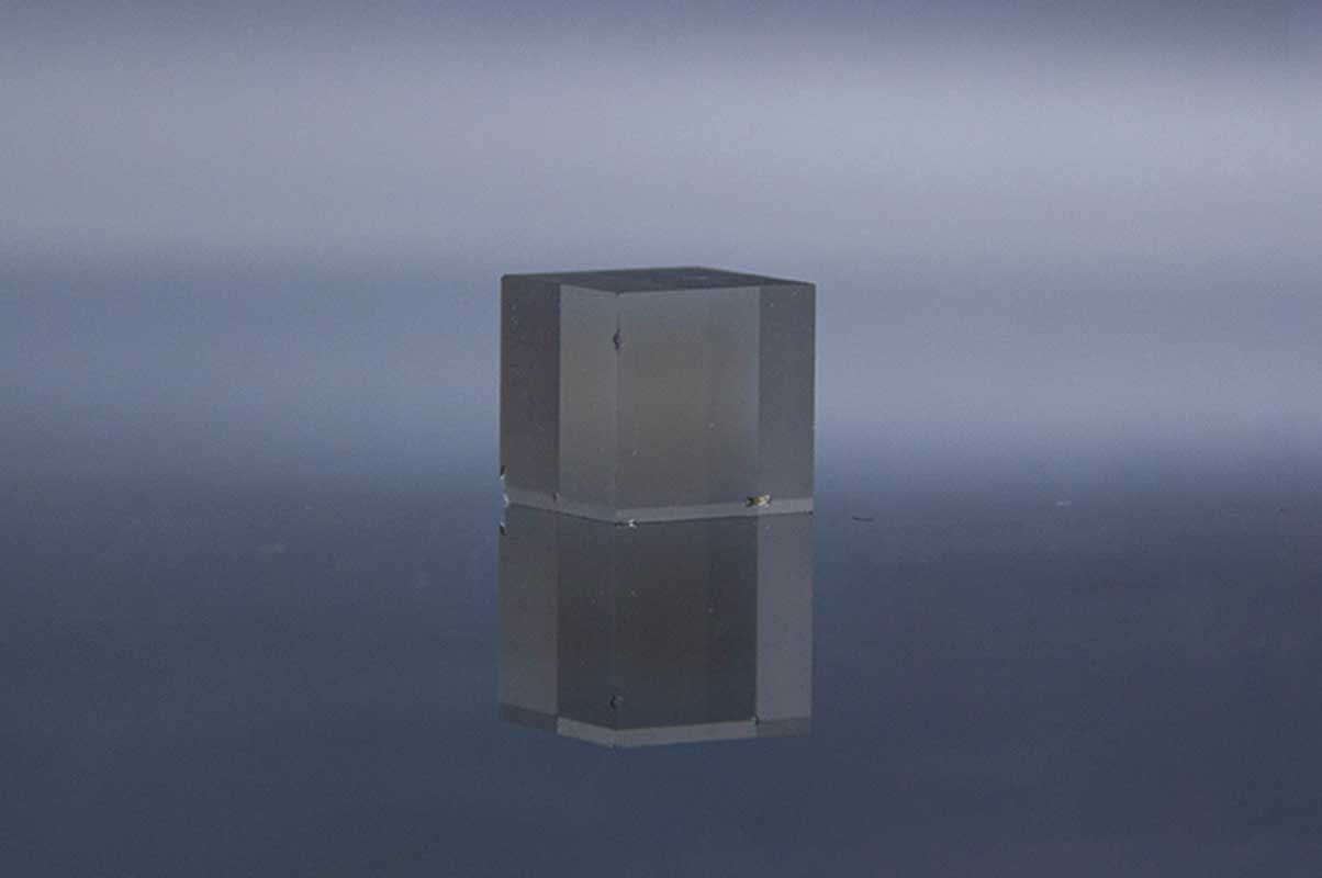
Ce:LuAG
| Wavelength (Max. Emission) (nm) | 535 |
| Wavelength range (nm) | 475-800 |
| Decay time (ns) | 70 |
| Light yield (Photons/kev) | 25 |
| Refractive index | 1.84@633nm |
The high resolution imaging system is composed of a high sensitivity digital CCD camera, an optical system and a thin scintillation screen. The screen may include Ce:LuAG inorganic scintillators.
[Ref. 1] The luminous efficiency (number of visible photons per keV) and spatial resolution of the two screens were compared. In the first experiment, the light detected by the CCD was averaged over a square ROI of 200μ 200 pixels in the center of the CCD. This Ce:YAG emission value of 17507 (the number of electrons in CCD pixels per second) and Ce:LuAG give a value of 26452, which is approximately Ce:YAG. This Ce:LuAG single crystal is more strongly absorbed by LuAG than yttrium aluminum garnet (density: 6.73 — 4.57 g/cm3) (the absorbed X-ray radiation (photon) in the range 1 to 40 keV is 1.7 times the attenuation coefficient of the X-ray radiation). The Ce:LuAG screen has a better SNR than the Ce:YAG screen for image application in imaging systems.
[Ref. 2] Images were taken using a Ce:LuAG 20 mm screen. The effective pixel size of CCD camera is 0.74mm. The operating voltage of the X-ray microfocusing source is 40kV/2mA. The image acquisition time was 5S, and 25 image samples were averaged. The measured intensity of the light produced by Ce:LuAG is about 1.51 times the Ce:YAG value after conversion of the photon flux emitted from the source. The light was examined with a CCD and averaged over a square ROI of 200×200 pixels. Compared with Ce:YAG (density: 6.73 — 4.57g/cm3), Ce:LuAG single crystal has a higher density, and LuAG has a stronger X-ray absorption (average X-ray absorption is 1.7 times). Range from 1 to 40keV).

References
[1] High-resolution application of YAG Ce and LuAG Ce imaging detectors with a CCD X-ray camera
[2] High-resolution imaging of biological and other objects with an X-ray digital camera
[3] Thin YAG Ce and LuAG Ce single crystal imaging plates used for high spatial resolution in X-ray imaging systems
Tl:CsI
| Wavelength (Max. Emission) (nm) | 410 |
| Decay time (ns) | 40 |
| Light yield (Photons/kev) | 25 |
| Light output relative to Nal (TI) (%) | 75 |
| Refractive index | 1.82@410nm |
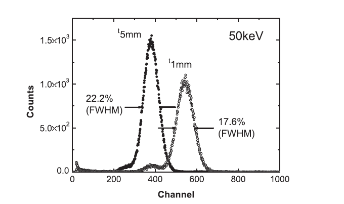
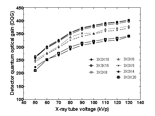
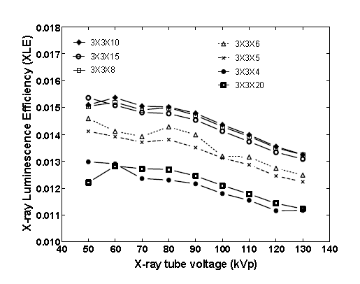
The most important characteristics of CsI(Tl) are its high optical yield (≥104 photons/MeV), and its emission spectrum has a maximum value at about 550 nm, which is fully compatible with amorphous and crystalline silicon photodiodes. The luminescence efficiency of CsI:Tl single crystal scintillation is studied as a function of the crystal thickness and X-ray tube voltage in the energy range used for mammography and general X-ray imaging.
[Ref. 1] Absolute luminous efficiency, which was experimentally determined for various X-ray energies used in X-ray imaging (40 — 140 kV) and mammography imaging (22 — 49 kV). Crystals were irradiated by X-ray using :(I) a philips X-ray setup with a tungsten anode target and a 2mm Al filter, and (ii) the general electronics Senographer DMR X-ray mammography setup with a molybdenum anode target and a molybdenum filter, using the experimental setup described previously.
[Ref. 2] To address these requirements, we developed a prototype fast X-ray imaging system using a structured CsI (TI) scintillator coupled to a fast frame 1K x 1K CCD. The system has been successfully used to capture 1024 x 64 pixel X-ray images at 1000 frames per second (FPS) with 12-bit dynamic range. The system goes beyond the capabilities of current high-speed X-ray imaging systems, which typically operate at 30 FPS. The high X-ray conversion efficiency of CsI (TI) (59,000 photons I MeV) makes these screens an excellent choice for current X-ray diffraction applications, where certain important diffraction peaks tend to be weak and require imaging screens with high SNR to be correctly identified.

References
[1] A systematic study of the performance of the CsI Tl single-crystal scintillator under X-ray excitation
[2] high-speed-xray-imaging-camera-using-structured-csitl-scintillator
[3] Scintillator efficiency study with MeV x-rays
[4] Structured CsI(Tl) scintillators for X-ray imaging applications
[5] Validation of columnar CsI x-ray detector responses obtained with hybridr , a CPU-GPU Monte Carlo code for coupled x-ray, electron, and optical transport
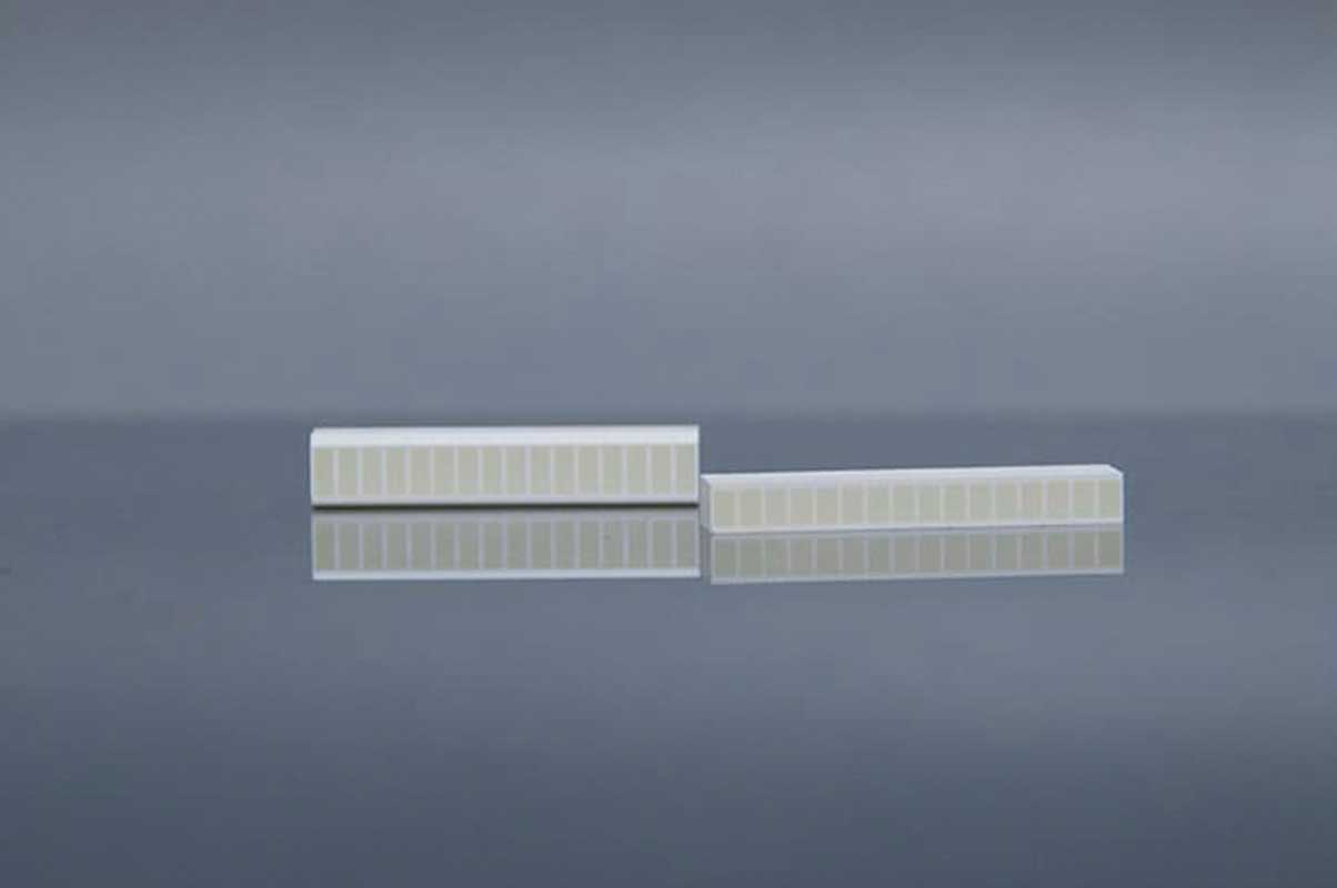
CdWO4
| Wavelength (Max. Emission) (nm) | 490 |
| Decay time (ns) | 14000 |
| Light yield (Photons/kev) | 12-15 |
| Light output relative to Nal (TI) (%) | 50 |
| Refractive index | 2.2-2.3 |
Cadmium tungstate (CdWO4, CWO) crystals are one of the candidates for such applications because of their high effective Z value, high density, higher light yield than BGO, and very low afterglow. The radiative hardness of the CWO crystal is up to 105 Rad. Because of this advantage, CWO scintillators are widely used in X-ray CT.
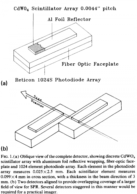
[Ref. 1] describes a high-resolution X-ray detector array for X-ray imaging consisting of a discrete array of CdWO4 scintillators and an Reticon 1024S photodiode array with fiber optic panels. It has developed a very high-resolution discrete scintillator array for use in fan-beam X-ray imaging systems.
References
[1] High-resolution digital x-ray detector utilizing a discrete array of CdWO4 scintillators and a self-scanned photodiode array
[2] Large Size CdWO4 Crystal for Energetic X- and γ Ray Detection
[3] X-RAY STUDY OF CHARACTERIZATION OF THE PbWO4 AND CdWO4 SINGLE CRYSTALS
GOS
| Wavelength (Max. Emission) (nm) | 510 |
| Decay time (us) | 5.5 |
| Relative light output (%) | 80 |
| Afterglow (%) | <0.01 |
| Luminescence intensity (keV) | 27.5 |
| GOS(Gd2O2S) | Density (g/cm3) | Wavelength of max emission (nm) | Decay constant (ns) |
| Ref[1] | 7.34 | 510 | 5.5μs |
References